Karnataka 1st PUC Biology Question Bank Chapter 8 Cell: The Unit of Life
1st PUC Biology Cell: The Unit of Life One Marks Questions and Answers
Question 1.
Who proposed the cell theory?
Answer:
Schleiden and Schwann.
Question 2.
Who discovered Nucleus?
Answer:
Robert Brown.
![]()
Question 3.
What is Pinocytosis?
Answer:
Ingestion of fluid material through plasma membrane is called Pincoytosis.
Question 4.
What is Phagocytosis?
Answer:
Ingestion of solid particules through plasma membrane is called phagocytosis.
Question 5.
What are the chemical components of the middle lamella?
Answer:
Calcium and magnesium pectate.
Question 6.
Who first used the term plastids?
Answer:
A.F.W. Schimper.
Question 7.
Who proposed fluid mosaic model of plasma membrane.
Answer:
S.J. Singer and G Nicolson.
![]()
Question 8.
What are plasmodesmata?
Answer:
The cytoplasmic connection between the cells is called plasmodesmata.
Question 9.
What is rough / smooth endoplasmic reticulum?
Answer:
Endoplasmic reticulum with ribosomes is called rough endoplasmic reticulum. Endoplasmic reticulum without ribosomes is called smooth endoplasmic reticulum.
Question10.
Name the membrane around Vacuole.
Answer:
Tonoplast.
Question 11.
Write two sub units of 70s ribosome / 80s ribosome.
Answer:
Sub units of 70s ribosome are 50s and 30s. Sub units of 80s ribosome are 60s and 40s.
Question 12.
What are Thylakoids?
Answer:
Membrane bound flattened sacs of granum are called Thylakoids.
Question 13.
Who discovered Lysosomes?
Answer:
Christian Duve:
Question 14.
Which cell organelle is also called ‘Suicide bag’ of cell?
Answer:
Lysosomes.
Question 15.
What are Ergastic substances?
Answer:
The non-living inclusions present in the cell cytoplasm are called Ergastic substances.
Question 16.
What are Raphides?
Answer:
Raphides are calcium oxalate crystals found in the vacuoles.
![]()
Question 17.
What is a cystolith?
Answer:
Grape like cluster of calcium carbonate crystals which hang from the certain palisade parenchyma cells of banyan leaf is cystolith.
Question18.
Name the power house of the cell.
Answer:
Mitochondria.
Question 19.
Who discovered Mitochondria?
Answer:
Kolliker.
Question 20.
Why is meiosis called a reduction division?
Answer:
Meiosis is called a reduction division because here the reduction in the number of chromosomes takes place. (Number of chromosomes reduces to half).
![]()
Question 21.
Name the site of protein synthesis.
Answer:
Ribosomes.
Question 22.
Which is the protein factory of the cell?
Answer:
Ribosome.
Question 23.
Mention any two functions of nucleus.
Answer:
(1) DNA present in the chromosomes is the primary hereditary material.
(2) Biosynthesis of DNX and its replication occurs in nucleus.
Question 24.
Which of the following is not correct?
(a) Robert Brown discovered the cell.
(b) Schleiden and Schwann formulated the cell theory.
(c) Virchow explained that cells are formed from pre-existing cells.
(d) A unicellular organism carries out its life activities within a single cell.
Answer:
(a) Robert Brown discovered the cell.
Question 25.
New cells generate from
(a) bacterial fermentation
(b) regeneration of old cells
(c) pre-existing cells
(d) abiotic materials
Answer:
(c) pre-existing cells
Question 26.
Which of the following is correct;
(a) Cells of all living organisms have a nucleus.
(b) Both animal and plant cells have a well defined cell wall.
(c) In prokaryotes, there are no membrane bound organelles.
(d) Cells are formed de novo from abiotic materials.
Answer:
(c) In prokaryotes, there are no membrane bound organelles.
Question 27.
What is a mesosome in a prokaryotic cell? Mention the functions that it performs,
Answer:
It is the infolding of plasma membrane found in prokaryotic cells.
Function of mesosomes are:-
(i) They help in cell wall formation, DNA replication and distribution to daughter cells.
(ii) They help in respiration, secretion process, to increase the surface area of the plasma membrane and enzymatic content.
Question 28.
Who gave the term cell?
Answer:
Robert Hooke (1665).
Question 29.
Who first observed the live cell?
Answer:
Anton Von Leewenhock (1677).
![]()
Question 30.
Who formulated the cell theory?
Ans.
M.J. Schleiden andT.Schwann (1838 – 39).
Question 31.
Expand PPLO?
Answer:
Pleuro Pneumonia like Organisms.
Question 32.
List out any four differences between plant cell and animal cell.
Answer:
Plaint cell: — Animal cell:
1. Cell wall is present. — Cell wall is absent.
2. Chloroplast is present. — Chloroplast is absent.
3. Centriole is absent. — Centriole is present.
4. Vacuoles are large. — Vacuoles are small.
Question 33.
What are plastids? Mention their types?
Answer:
Pigment containing cell organelle found in plant cells are called plastids. They are three types.
(i) Chloroplast (green).
(ii) Chromoplast (coloured) and
(iii) Leucoplast (colorless).
Question 34.
Differentiate between gram positive and gram negative bacteria.
Answer:
| gram positive | gram negative |
| 1. They are stained by gram 1 stain | 1. They are not stained by gram stain. |
| 2. Cell wall is thin. | 2. Cell wall is thick. |
1st PUC Biology Cell: The Unit of Life Two Marks Questions and Answers
Question 1.
List any four functions of the cell membrane.
Answer:
(i) Cell membranae is a selectively permeable membrane so that, it permits water and other selected materials to get in or out.
(ii) It helps in Pinocytosis.
(iii) It helps in Phagocytosis.
(iv) It helps in the absorption of ions and molecules. (Active transport).
![]()
Question 2.
Multicellular organisms have division of labour. Explain.
Answer:
Multicellular organisms possess tissues, organs and organ systems which perform diverse functions like digestion, respiration, excretion, circulation, etc. Thus they exhibit division of labour.
Question 3.
Both lysosomes and vacuoles are endomembrane structures, yet they differ in terms of their functions. Comment.
Answer:
Lysosomes:
Lysosomes are single member saclike structures found in most of the eukaryotic cells but absent in prokai yotes. membrane is lipoproteinaceous in nature. (Absent in 0R13C’s. yeast and Eiiglcna) Cheletc’ and corticosterols give stability to the membrane and prevent damages by enzyme. [he matrix of the lysosome contains hydrolytic and lipolytic enzymes.
Lysosomes are regarded as ‘suicide bags’ as they take part in intracellular digestion, especially when the cell experiences starved condition or acute problem of survival.
Lysosomes show polymorphism and exist in four types as follows:
1. Primary lysosomes: These are formed by the Golgi Complex and contain enzymes synthesized by Rough ER. They give rise to secondary lysosomes.
2. Secondary lysosomes: These are formed from primary lysosomes and these are also known as digestive vacuoles or heterophagosomes. They take part in phagocytosis (engulfing bacteria and other foreign invaders) and pinocytosis (drinking water impurities in cytoplasm).
3. Tertiary lysosomes: The indigestible or undigested material present in secondary lysosome forms materials of tertiary lysosomes which are known as Residual bodies. Tertiary lysosomes also take part in intracellular digestion of pathogens.
![]()
4. Autophagosomes/Autolysosomes: These differentiate from primary Tysosomes (seen in mitochondria and ER) and are self motivated to engulf the cellular contents i.e. self digestion or autolysis in case of deficiency of food.
Vacuole:
These are non cytoplasmic areas, present inside the cytoplasm and bounded by a single layer of semiperneable membrane called Tonoplast.
Vacuoles mainly occur in Eukaryotes. in animal cells, the size of the vacuole is smaller and in plant cells, the size is larger and is central in position.
Functions:
Based on functions, vacuoles are of the following types:
(a) Sap vacuoles: Present in plant cells which store water and dissolved mineral ions. They function as osmometers.
(b) Contractile vacuoles: Present in protozoans like amoeba, paramaecium etc., help in osmoregulation and excretion.
(c) Food vacuoles: Present in protozoans and help in the digestion of food substances.
(d) Gas vacuoles: Present in protozoans, contain gases like O2, CO2, N2 etc., and provide buoyancy.
Question 4.
List any four functions of endoplasmic reticulum.
Answer:
Endoplasmic reticulum is a well developed electron microscopic network of double membrane bound channels distributed through out the cytoplasm extending from the nucleus to the margins of the cell (more concentrated in the endoplasm portion of the cytoplasm). ER is fully developed and differentiated in Eukaryotic cells, but absent in prokaryotes.
Morphologically the ER is composed, of the following three kinds of structures. Namely.
(a) Cisternae
(b) Vesicles
(c) Tubules.
- Cisternae are long flattened sac like unbranched tubules of 40-50 microns diameter arranged parallely in bundles.
- Vesicles are oval, membrane bound vacuolar structures, scattered in the cytoplasm. Their diameter ranges from 25 to 500 microns.
- Tubules are wider, tubular, branched elements of 50 to 190 microns in diameter. All these three different types of components join in a reticulate manner to form the ‘cytoskeleton’ of the ER.
- The ER remains continuous with the membranes of plasma membrane nuclear membrane and Golgi complex.
![]()
Based on the presence or absence of ribosomes, the ER is of two types:
1. Smooth Endopbsmic Reticulum:-
- Without ribosomal particles.
- Present mainly near cell (membrane.
- Mainly consists of tubules.
- Formed from rough ER by the loss of ribosomes.
2. Rough ER (Granular ER):
- Ribosomes present on the surface of the ER.
- Mainly present near the nucleus.
- Consists of cisternae.
- Formed from nuclear membrane.
Question 5.
Classify lysosomes based on their functions?
Answer:
Functions:
1. They are seen in Acrosome of’ sperms, where they release enzymes which aid help in fertilization (by dissolving the egg wall).
2. They are involved in destruction of cells of the tail in tadpole during its metamorphosis to form an adult tadpole.
3. They take part in septic reactions by mediating irritation and pain.
4. They are seen in lymphoid systems, and help in the destruction or break down of dead cells and pathogens.
5. Lysosomes break down old cell organdies to their respective molecular components for resynthesis of organelles. Thus they help in cell recycling.
6. During starvation and disease lysosomes digest lipids, proteins and glycogen to supply energy to the body. This is called intra cellular digestion of macro molecules.
![]()
Question 6.
List the function of golgi complex.
Answer:
Golgi complex is regarded as the enzyme factory of the cell. The golgi complex is regarded as golgi material and golgi bodies in eukaryotic advanced animals, and known as dictyosomcs in plants and lower animals. Generally golgi complex is absent in prokaryotic cells.
It is generally abundant in secretory cells. Golgi Complex is made Lip of double membrane flattened sacs called cisternae, associated with peripherally occurring large vacuolar vesicles and network of interconnecting tubules. It appears to be continuous with E.R and thus regarded to be derived from it.
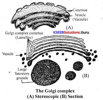
Functions:
1. It secretes various enzymes and hormones.
2. It forms secretory vesicles which store enzymes, proteins, carbohydrates etc.
3. It takes part in the formation of cell plate and cell wall along with ER at the time of
telophase in plant cells.
4. It helps in the formation of primary lysosomes.
5. It forms the acrosome at the sperms.
6. It is involved in-cell secretion.
e.g: Polysaccharides in goblet cells of intestine (which form mucous which lubricates the intestine during digestion of food).
Question 7.
What are ergastic substances? Give two examples.
Answer:
The non-living inclusions of the cell are called Ergastic substances. These are either products of metabolism or by-products which are of varied nature. They occur in cytoplasm, vacuole or in cell wall. They may be classified into:
(a) Reserved food materials
(b) Secretory products
(c) Excretory products
(d) Mineral crystals
(a) Reserve food materials: The reserve food materials in the cell includes starch grains,protein grains, oil globules in plant cells and glycogen granules in the animal cell.
![]()
(b) Secretory products: These are chemical compounds secreted by the cytoplasm or cell organdies. They include enzymes, hormones, pigments like chiorophylls, carotene and xanthophylls in plants and haemoglobin in animals and Nector (produced by nectaries in flowers).
(c) Excretory products: Most of the waste products are formed due to metabolic activities in the plant body and are commercially important materials. They accumulate in leaves,fruits, bark and sometimes in the roots of the plants.
1st PUC Biology Cell: The Unit of Life Three Marks Questions and Answers
Question 10.
Match the following Column I
Column I — Column II
(a) Cristae — (i) Flat membranous sacs in stroma.
(b) Cisternae — (ii) Infoldings in mitochondria.
(c) ThyIakoids — (iii) Disc-shaped sacs in golgi apparatus.
Answer:
(a) (ii), (b) (iii), (c) (i).
1st PUC Biology Cell: The Unit of Life Five Marks Questions and Answers
Question 1.
Draw a neat labelled diagram of the ultrastructure of a typical animal cell.
Answer:
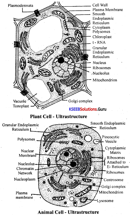
![]()
Question 2.
Describe the structure of nucleus with a neat labelled diagram.
Answer:
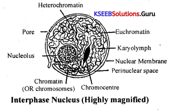
The nucleus is a dynamic celt organelle which actively and randomly controls the functioning of all other cell organelles either electrochemically or neurochemically. The shape of the nucleus varies in different cells. Normally it is spherical, but it may be oval, discoid, kidney shaped or lobed. The size of the nucleus is also variable. Generally there is a single nucleus in a cell, but’ some cells also contain two or more nuclei.
The nucleus is composed of following components:
(a) Nuclear membrane: The nucleus is bounded by a double layered nuclear membrane with pores at intervals. The space between the two membranes is known as the per nuclear space.
(b) Nucleoplasm: Within the boundary of nuclear membrane, a specialized cytoplasm is present known as nucleoplasm or karyoplasm, which forms the matrix. Bathed in the nucleoplasm are present the nucleolus and chromosomes.
(c) Nucleolus: The nucleolus is a spherical dark body, seen in contact with a specific chromosome at a point called nucleolar organizer. The peripheral portion of the nucleolus’ is amorphous and contains RNA. The central portion is crystalline and contains DNA. It controls the activities of nucleus and other cell organdIes and thus regarded as the, ‘Nucleus of the nucleus’.
![]()
(d) Chromosomes: Chrómosomes are the thread-like coloured bodies that are in the intranuclear position which act as the vehicles of heredity and variations. They are self-replicating, and exhibit cyclic change in size and shape. They are visible during cell division; Waldeyer described them in 1888. Chromosomes are the self reproducing components of nucleus with DNA, which become visible during cell division. They exhibit cyclic change in size & shape.
Question 3.
How do neutral solutes move across the plasma membrane? Can the polar molecules also move Across it, in the same way? If not, then how are these transported across the membrane?
Answer:
Neutral solutes move across process of simple diffusion or osmosis. Polar molecules move by facilitated diffusion, or active transport through carriers or protein pumps.
Question 4.
Name two cell organelles that are double membrane bound. What are the characteristics of these two organelles? State their functions and draw labelled diagrams of both.
Answer:
Double membrane cell organelles are plastids (e.g., chloroplasts) and mitochondria.
Modifications of plasma membrane.
To perform specific functions and exhibit flexibility, PM undertakes the following modifications:
1. Microvilli: These are minute folding of plasma membrane to increase surface area for absorption.
2. Desmosomes: Inner surface of adjacent plasma membranes have thickened areas called desmosomes which help in cell adhesion.
3. Pinocytic vesicies: Invaginations of the plasma-membrane into cytoplasm and fomation of upficlds which contain fluids.
4. Mesosomes: In prokaryotes, invaginations of the plasma membrane are associated with the respiratory system enzymes and help in respiration and are called as mesosomes.
The functions of the plasma membrane are:
1. It maintains the size and sbape of the cell.
2. Osmosis: Osmosis is the process by which water molecules pass through a semi permeable membrane (here it is the plasma membrane) from the region of its higher concentration- to one of lower concentration.
3. Active transport: It is energy (from ATP) dependent transport of molecules or ions across a semi permeable membrane against the concentration (electrochemical gradient). It can also be called “metabolically linked transport”. e.g: Sodium – Potassium Pump: (Revolving door model) :
The moving machinery of Na+ & K+ through active transport is called ‘Sodium – Potassium pump’.
Question 5.
What are the characteristics of prokafyotic cells.
Answer:
Characteristics of Prokaryotic cell are as follows.
(i) Prokaryotic cells are smaller.
(ii) They have cell wall, cell membrane and cytoplasm. They do not have a well defined nucleus.
(iii) Few prokaryotes like bacteria have an additional small circular DNA called plasmids.
(iv) Prokaryotic cell does not contain membrane bound organelles as found in eukaryotes except ribosomes.
(v) A specialised differentiated form of cell membrane called mesosonie is found in prokaryotes.
![]()
Question 6.
Describe the structure of the following with the help of labelled diagrams.
(i) Nucleus
(ii) Centrosome
Answer:
The nucleus is a dynamic celt organelle which actively and randomly controls the functioning of all other cell organelles either electrochemically or neurochemically. The shape of the nucleus varies in different cells. Normally it is spherical, but it may be oval, discoid, kidney shaped or lobed. The size of the nucleus is also variable. Generally there is a single nucleus in a cell, but’ some cells also contain two or more nuclei.
The nucleus is composed of following components:
(a) Nuclear membrane: The nucleus is bounded by a double layered nuclear membrane with pores at intervals. The space between the two membranes is known as the per nuclear space.
(b) Nucleoplasm: Within the boundary of nuclear membrane, a specialized cytoplasm is present known as nucleoplasm or karyoplasm, which forms the matrix. Bathed in the nucleoplasm are present the nucleolus and chromosomes.
(c) Nucleolus: The nucleolus is a spherical dark body, seen in contact with a specific chromosome at a point called nucleolar organizer. The peripheral portion of the nucleolus’ is amorphous and contains RNA. The central portion is crystalline and contains DNA. It controls the activities of nucleus and other cell organdIes and thus regarded as the, ‘Nucleus of the nucleus’.
![]()
(d) Chromosomes: Chrómosomes are the thread-like coloured bodies that are in the intranuclear position which act as the vehicles of heredity and variations. They are self-replicating, and exhibit cyclic change in size and shape. They are visible during cell division; Waldeyer described them in 1888. Chromosomes are the self reproducing components of nucleus with DNA, which become visible during cell division. They exhibit cyclic change in size & shape.
This structure ¡s a conspicuous structure in animal cells (except RBC’s). Absent in prokaryotes and higher plants. Centrosome is present near the nucleolus, Centrosome has two darkly stained cylindrical
bodies called centrioles at right angles to each other. These are seen by transparent cytoplasmic area called centrosphere. Since the centrioles are in pairs, they are called Diplosome.
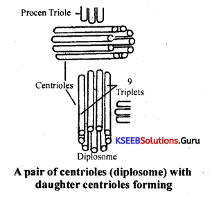
Ultra structure: Each of the centriole is made up of 9 sets of microtubules. Each of this microtubules is made up of three sùbtubules (triplet) which inturn are made up of globular proteins. The microtubu les are connected to each other the by dense material
strand.
Functions:
1. At end of prophase of mitosis in animal cells, centriole duplicate and new daughter centrioles are lormed.
2. Give rise to ñasal bodies from which cilia and flagella arise.
3. Distal centriole of sperm gives rise to axonemes or axial filament of sperm tail.
Question 7.
What is a centromere? How does the position of centromere form the basis of classification of chromosomes. Support your answer with a diagram showing the position of centromere on different types of chromosomes.
Answer:
This structure ¡s a conspicuous structure in animal cells (except RBC’s). Absent in prokaryotes and higher plants. Centrosome is present near the nucleolus, Centrosome has two darkly stained cylindrical
bodies called centrioles at right angles to each other. These are seen by transparent cytoplasmic area called centrosphere. Since the centrioles are in pairs, they are called Diplosome.
![]()
Ultra structure: Each of the centriole is made up of 9 sets of microtubules. Each of this microtubules is made up of three sùbtubules (triplet) which inturn are made up of globular proteins. The microtubu les are connected to each other the by dense material
strand.
Functions:
1. At end of prophase of mitosis in animal cells, centriole duplicate and new daughter centrioles are lormed.
2. Give rise to ñasal bodies from which cilia and flagella arise.
3. Distal centriole of sperm gives rise to axonemes or axial filament of sperm tail.
Question 8.
Explain the structure of mitochondria with a neat labelled diagram.
Answer:
It is present in eukaryotes except mammalian RBC, and absent in prokaryotes. Its shape is oval, or sausage. Its number per cell depends on the metabolic state of the cell.
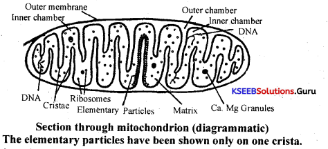
Structure: The mitochondria is bounded by two lipoprotein unit membranes namely outer membrane and inner membrane. In between them, lies perimitochondrial matrix containing water, minerals and enzymes.
Outer membrane is smooth and unfolded while the inner membrane is folded and it produces inward finger like process called Cristae. Along the inner surface of the inner membrane, there are numerous tiny tadpole like structures called elementary particles or F1 particles, Racker’s particles or oxysomes. F1 particle contains basal piece, stalk and head. Sites of ATPase is between adjacent elementary particles.
The inner membrane contains electron transport system. It is made up of a chain of co-enzymes in the order of NAD, FAD, cytochrome B, cyt C, cyt A, cyt A3. The inner space of mitochondria is filled with a dense fluid called mitochondria matrix containing water, proteins, lipids, all enzymes of Kreb’s cycle, circular DNA and 80s ribosomes.
Functions:
I. Mitochondria are the centers of aerobic respiration.
2. They are the sites of synthesis and storage of energy as ATP. Hence called power house of the cell.
3. As they have circular DNA & ribosomes, they synthesise a few proteins for their own requirement. Hence they are called ‘Semi autonomous cell organeíles’.
Question 9.
Explain the structure of chloroplast with a neat labelled diagram.
Answer:
Chlorophyll containing plastids are called Chioroplasts. They are present in the cells of all green plants and abundant in the leaf mesophyll cells. They are elliptical or oval in shape.
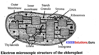
Chloroplast is bounded by two membranes with the inter membrane space called peri plastidal space, containing peri plastidal fluid. It is made up of H2O, mineral ions, proteins etc. It is a lubricating fluid which avoids friction between the two membranes. The inner chamber is filled with a colourless proteinaceous fluid matrix called stroma. Besides abundant proteins, stroma contains 70s ribosomes, circular DNA and all the enzymes of the Calvin cycle.
![]()
Embedded in the stroma, there are green coloured bodies called grana, which are interconnected by frets. Grana is the site of light reaction, each granum consists of a stack of lipoprotein membrane discs called Thylakoids. Each thylakoid contains several photosynthetic centers called quantasomes. Each quantasome contains about 250 chlorophyll pigments and a few xanthophylls and carotenes.
Only chlorophyll- a is capable of harvesting light energy into photosynthesis and hence it is called the primary photosynthetic pigment. All the other pigments, merely absorb light energy and pass it on to chlorophyll, and hence are called accessory photosynthetic pigments.
Note: As chloroplasts contains circular DNA 70s ribosomes and as they go for protein synthesis, they are regarded as semi autonomous cell organelles.
1st PUC Biology Cell: The Unit of Life Text Book Questions and Answers
Cell Structure:
The study of cells is called Cell Biology or Cytology. Robert Hook used the term cell first. In 1838 Schleiden and Schwann proposed cell theory. According to cell theory:
1. All living things are made up of protoplasmic units called cells.
2. All cells are basically alike, both in structure and metabolic functions.
General structure of the cell:
A cell basically contains a nucleus and cytoplasm within a limiting membrane brown as the cell membrane. Each cell has a definite age (life span) and performs various metabolic activities (respiration, digestion, excretion, reproduction etc).
Any organism i.e. simple (unicellular) or complex (multicellular) is nothing but an amplified version of the cellular functions. Thus, cell can be regarded as the structural and functional basis of life.
A cell, apart from nucleus and cytoplasm contains various other living and non-living components in it. The living ‘components are all termed as organelles and these components are in perfect harmony, within the cell.
![]()
Living Components:
1. Mitochondria (Power house).
2. Plastids (antennae).
3. Lysosomes (suicide bags).
4. Centrosomes (cell division apparatus).
5. Golgi complex (enzyme factory).
6. Endoplasmic reticulum (skeletal network).
7. Ribosomes (protein factory).
Non-living inclusions:
1. Reserve food materials.
3. Excretory materials.
5. Vacuole.
Cell Wall:
It is outer most rigid, non-living structure of a plant cell. It is a characteristic of metaphyta plant cells. It is also found in fungi, bacteria, cyanobacteria and some protista members. Cell wall is normally made up of two layers:
(a) Primary wall: It is the initial wall, which is the first secretion product of the cytoplasm. It is thin and elastic, and made up of pectin and cellulose. Primary wall of adjacent cells are cemented by the middle lamellae i.e., in between two primary walls. The cementing properties is due to the presence of pectin and Mg, Ca pectates which give rigidness to the cell wall.
(b) Secondary cell wall: It is formed by depositions on the inner surface of a primary wall. It has 3 layers viz, outer S1, middle S2 and inner S3 in a concentric manner. It may vary in hardness, thickness and colour. This forms the wood and fibres. It is mainly made up of cellulose, hemicellulose and later may be deposited with suberin, cutin or lignin occasionally.
(c) Tertiary cell wall: Occasionally a tertiary cell wall may be deposited in some cells within the secondary cell wall which are chemically made up of xylan.
Plasmodesmata: The cytoplasm of one cell is connected with that of the adjoining cell by fine cytoplasmic strands which extend through extremely minute pits that are left in the cell wall during its formation. These cytoplasmic strands are called plasmodesmata.
Functions:
1. The cell wall is dead and it gives physical, chemical‘and biological protection.
2. It is permeable to various substances required by cells.
3. It provides definite shape, mechanical strength to its the cell due to thick, rough and tough nature.
4. It bears pits through which plasmodesmata extend, which helps in the movement of substances from one cell to the other.
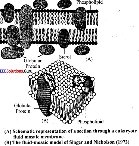
All living cells are enclosed in a thin, living and selectively permeable membrane called plasma membrane. Its thickness is about 75 – 100 A Plasma membrane consists of 30 – 40% lipids and 60 – 70% proteins. In animal cells, it surrounds the protoplasm and in plant cell it is next to cellwall.
Fluid mosaic Model By Singer & Nicholson:
a. According to this model, the plasma membrane consists of a double layer of lipid molecules and globular protein molecules and sterols distributed at random. In comparative term it can be said that the plasma membrane is formed of ‘protein icebergs in the sea of lipids’.
b. The protein molecules are globular proteins and these proteins penetrate or lie at the periphery to form a mosaic pattern.
c. Heads of the phospholipid molecules of the two layers are directed in the opposite directions while tails of the two layers face each other.
d. In animal cells glycolipids and cholesterol are also present along with proteins.
![]()
Modifications of plasma membrane.
To perform specific functions and exhibit flexibility, PM undertakes the following modifications:
1. Microvilli: These are minute folding of plasma membrane to increase surface area for absorption.
2. Desmosomes: Inner surface of adjacent plasma membranes have thickened areas called desmosomes which help in cell adhesion.
3. Pinocytic vesicles: Invaginations of the plasma-membrane into cytoplasm and fomation of upfields which contain fluids.
4. Mesosomes: In prokaryotes, invaginations of the plasma membrane are associated with the respiratory system enzymes and help in respiration and are called as mesosomes.
The functions of the plasma membrane are:
1. It maintains the size and shape of the cell.
2. Osmosis: Osmosis is the process by which water molecules pass through a semi permeable membrane (here it is the plasma membrane) from the region of its higher concentration- to one of lower concentration.
3. Active transport: It is energy (from ATP) dependent transport of molecules or ions across a semi permeable membrane against the concentration (electrochemical gradient). It can also be called “metabolically linked transport”.
e. g: Sodium – Potassium Pump: (Revolving door model) :
The moving machinery of Na+ & K+ through active transport is called ‘Sodium – Potassium pump’.
Cytoplasm:
It is an amorphous, translucent, homogenous colloidal substance lying between plasma membrane and nuclear membrane. It is divided into outer ectoplasm and inner endoplasm.
Functions:
1. It provides raw materials to various cell organelles for their functioning and helps in the exchange of materials.
2. It is the seat of biosynthesis of organic biomolecules like fats, nucleotides, proteins etc, and also catabolism. It is also the seat for other biological activities like reproduction, growth, irritability etc.
Mitochondria: (Power house of Cell)
It is present in eukaryotes except mammalian RBC, and absent in prokaryotes. Its shape is oval or sausage. Its number per cell depends on the metabolic state of the cell.
Structure: The mitochondria is bounded by two lipoprotein unit membranes namely outer membrane and inner membrane. In between them, lies perimitochondrial matrix containing water, minerals and enzymes.
Outer membrane is smooth and unfolded while the inner membrane is folded and it produces inward finger like process called Cristae. Along the inner surface of the inner membrane, there are numerous tiny tadpole like structures called elementary particles or F1 particles, Racker’s particles or oxysomes. F1 particle contains basal piece.
stalk and head. Sites of ATPase is between adjacent elementary particles. The inner membrane contains electron transport system. It is made up of a chain of co-enzymes in the order of NAD, FAD, cytochrome B, cyt C, cyt A, cyt A3. The inner space of mitochondria is filled with a dense fluid called mitochondria matrix containing water, proteins, lipids, all enzymes of Kreb’s cycle, circular DNA and 80s ribosomes.
Functions:
1. Mitochondria are the centers of aerobic respiration.
2. They are the sites of synthesis and storage of energy as ATP. Hence called power house of the cell. .
3. As they have circular DNA & ribosomes, they synthesise a few proteins for their own requirement. Hence they are called ‘Semi autonomous cell organelles’.
Plastids:
Plastids are oval shaped or disc shaped structures present in algae and green plants.
(1) Leucoplasts
(2) Chloroplasts
(3) Chromoplasts.
1. Leucoplast: These plastids are without pigments (Colour less plastids). They are useful in storage of fats starch and proteins, which are synthesized products. They occur in storage roots, stems, leaves & parts of the plant not exposed to light
2. Chromoplasts: These are plastids which contain coloured pigments except the green pigment chlorophyll.
They are sub divided into:
- Blue green chromoplasts: Phycocyanin is the main pigment.
- Pheoplasts: Main pigments are Phycoxanthin and other Xanthophylls.
- Rhodoplasts: Phycoerythrin is the main pigment.
![]()
3. Chloroplast:
Chlorophyll containing plastids are called Chloroplasts. They are present in the cells of all green plants and abundant in the leaf mesophyll cells. They are elliptical or oval in shape. Chloroplast is bounded by two membranes with the inter membrane space called peri plastidal space, containing peri plastidal fluid. It is made up of H2O, mineral ions, proteins etc. It is a lubricating fluid which avoids friction between the two membranes.
The inner chamber is filled with a colourless proteinaceous fluid matrix called stroma. Besides abundant proteins, stroma contains 70s ribosomes, circular DNA and all the enzymes of the Calvin cycle. Embedded in the stroma, there are green coloured bodies called grana, which are inter connected by frets. Grana is the site of light reaction, each granum consists of a stack of lipoprotein membrane discs called Thylakoids. Each thylakoid contains several photosynthetic centers called quantasomes. Each quantasome contains about 250 chlorophyll pigments and a few xanthophylls and carotenes.
Only chlorophyll- a is capable of harvesting light energy into photosynthesis and hence it is called the primary photosynthetic pigment. All the other pigments, merely absorb light energy and pass it on to chlorophyll, and hence are called accessory photosynthetic pigments.
Note: As chloroplasts contains circular DNA 70s ribosomes and as they go for protein synthesis, they are regarded as semi autonomous cell organelles.
Endoplasmic Reticulum:
Endoplasmic reticulum is a well developed electron microscopic network of double membrane bound channels distributed through out the cytoplasm extending from the nucleus to the margins of the cell (more concentrated in the endoplasm portion of the cytoplasm). ER is fully developed arid differentiated in Eukaryotic cells, but absent in prokaryotes.
Morphologically the ER is composed of the following three kinds of structures. Namely,
(a) Cisternae
(b) Vesicles
(c) Tubules.
• Cisternae are long flattened sac like unbranched tubules of 40-50 microns diameter arranged parallely in bundles.
• Vesicles are oval, membrane bound vacuolar structures, scattered in the cytoplasm. Their diameter ranges from 25 to 500 microns.
• Tubules are wider, tubular, branched elements of 50 to 190 microns in diameter.
All these three different types of components join in a reticulate manner to form the ‘cytoskeleton’ of the ER.
The ER remains continuous with the membranes of plasma membrane nuclear membrane and Golgi complex.
Based on the presence or absence of ribosomes, the ER is of two types:
1. Smooth Endoplasmic Reticulum:-
• Without ribosomal particles.
• Present mainly near cell, membrane.
• Mainly consists of tubules.
• Formed from rough ER by the loss of ribosomes.
2. Rough ER (Granular ER):-
• Ribosomes present on the surface of the ER.
• Mainly present near the nucleus.
• Consists of cisternae.
• Formed from nuclear membrane.
Functions:-
Serves as secretary, storage, circulatory and nervous system for the cell).
1. ER forms the ultra structural skeletal frame work (cytoskeleton) to the cell and gives mechanical support to the colloidal cytoplasmic matrix.
2. It takes part in transportation of substances. It is an efficient intracellular transportation channel which connects nuclear membrane and cell membrane and permits the exchange of molecules through its membranes by the process of osmosis, diffusion and active transport.
3. It provides increased surface area of various enzymatic reactions like protein synthesis. Also, it contains many enzymes, and thus takes part in synthetic and metabolic activities.
4. It is useful in intracellular impulse- conduction, and thus nucleus is able to respond quickly to change in the cell’s immediate environment.
5. It forms the new nuclear envelop, after each nuclear division.
6. It contributes to the formation of cell plate, during telophase of cell division in plants.
7. Smooth endoplasmic reticulum takes part in lipid synthesis, break down of glycogen and detoxification of drugs.
8. Rough endoplasmic reticulum takes part in protein synthesis and secretion of enzymes.
Golgi Complex:
Golgi complex is regarded as the enzyme factory of the cell. The golgi complex is regarded as golgi material and golgi bodies in eukaryotic advanced animals, and known as dictyosomes in plants and lower animals. Generally golgi complex is absent in prokaryotic cells.
It is generally abundant in secretory cells. Golgi Complex is made up of double membrane flattened sacs called cisternae, associated with peripherally occurring large vacuolar vesicles and network of inter connecting tubules. It appears to be continuous with E.R and thus regarded to be derived from it.
Functions:
1. It secretes various enzymes and hormones.
2. It forms secretory vesicles which store enzymes, proteins, carbohydrates etc.
3. It takes part in the formation of cell plate and cell wall along with ER at the time of telophase in plant cells.
4. It helps in the formation of primary Iysosomes.
5. It forms the acrosome at the sperms.
6. It is involved in-cell secretion.
e.g: Polysaccharides in goblet cells of intestine (which form mucous – which lubricates the intestine during digestion of food).
![]()
Centrosome:
This structure is a conspicuous structure in animal cells (except RBC’s). Absent in prokaryotes and higher plants.
Centrosome is present near the nucleolus. Centrosome has two darkly stained cylindrical bodies called centrioles at right angles to each other. These are seen by transparent cytoplasmic area called centrosphere. Since the centrioles are in pairs, they are called Diplosome.
Ultra structure: Each of the centriole is made up of 9 sets of microtubules. Each of this microtubules is made up of three subtubules (triplet) which inturn are made up of globular proteins. The microtubules are connected to each other the by dense material strand.
Functions:
1. At end of prophase of mitosis in animal cells, centriole duplicate and new daughter centrioles are formed.
2. Give rise to basal bodies from which cilia and flagella arise. ’
3. Distal centriole of sperm gives rise to axonemes or axial filament of sperm tail.
Lysosomes:
Lysosomes are single membrane saclike structures found in most of the eukaryotic cells but absent in prokaryotes. Tbs membrane is lipoproteinaceous in nature. (Absent in RBC’s, yeast ana Euglena) Cholesterol and corticosterols give stability to the membrane and prevent damages by enzymes. The matrix of the lysosome contains hydrolytic and lipolytic enzymes.
Lysosomes are regarded as ‘suicide bags’ as they take part in intracellular digestion, especially when the cell experiences starved condition or acute problem of survival.
Lysosomes show polymorphism and exist in four types as follows:
1. Primaiy lysosomes: These are formed by the Golgi Complex and contain enzymes synthesized by Rough ER.
They give rise to secondary lysosomes.
2. Secondary lysosomes: These are formed from primary lysosomes and these are also
known as digestive vacuoles or heterophagosomes. They take part in phagocytosis (engulfing bacteria and other foreign invaders) and pinocytosis (drinking water impurities in cytoplasm). .
3. Tertiary lysosomes: The indigestible or undigested material present in secondary lysosome forms materials of tertiary lysosomes which are known as Residual bodies. Tertiary lysosomes also take part in intracellular digestion of pathogens.
4. Autophagosomes/Autolysosomes: These differentiate from primary lysosomes (seen in mitochondria and ER) and are self motivated to engulf the cellular contents i.e. self digestion or autolysis in case of deficiency of food.
Functions:
1. They are seen in Acrosome of sperms, where they release enzymes which aid help in fertilization (by dissolving the egg wall).
2. They are involved in destruction of cells of the tail in tadpole during its metamorphosis to form an adult tadpole.
3. They take part in septic reactions by mediating irritation and pain.
4. They are seen in lymphoid systems, and help in the destruction or break down of dead cells and pathogens.
5. Lysosomes break down old cell organelles to their respective molecular components for resynthesis of organelles. Thus they help in cell recycling.
6. During starvation and disease lysosomes digest lipids, proteins and glycogen to supply energy to the body. This is called intra cellular digestion of macro molecules.
![]()
Nucleus:
The nucleus is a dynamic ceil organelle which actively and randomly controls the functioning of all other cell organelles either electrochemically or neurochemically.
The shape of the nucleus varies in different cells. Normally it is spherical, but it may be oval, discoid, kidney shaped or lobed. The size of the nucleus is also variable. Generally there is a single nucleus in a cell, but-some cells also contain two or more nuclei.
The nucleus is composed of following components:
(a) Nuclear membrane: The nucleus is bounded by a double layered nuclear membrane with pores at intervals. The space between the two membranes is known as the per nuclear space’.
(b) Nucleoplasm: Within the boundary of nuclear membrane, a specialized cytoplasm is present known as nucleoplasm or karyoplasm, which forms the matrix. Bathed in the nucleoplasm are present the nucleolus and chromosomes.
(c) Nucleolus: The nucleolus is a spherical dark body, seen in contact with a specific chromosome at a point called nucleolar organizer. The peripheral portion of the nucleolus is amorphous and contains RNA. The central portion is crystalline and contains DNA. It controls the activities of nucleus and other cell organelles and thus regarded as the ‘Nucleus of the nucleus’.
(d) Chromosomes: Chrdmosomes are the thread-like coloured bodies that are in the intranuclear position which act as the vehicles of heredity and variations. They are self-replicating, and exhibit cyclic change in size and shape. They are visible during cell division. Waldeyer described them in 1888.
Chromosomes are the self reproducing components of nucleus with DNA, which become visible during cell division. They exhibit cyclic change in size & shape.
![]()
1. Chromosome size: The size of the chromosome is measured at the metaphase of mitosis. It is constant for a particular species. Its length varies from 0.2 to 50 p. Diameter 0. 2 to 2p. Chromosomes of plants are larger than that of animals. Monocots have very large ones. More the number of chromosomes per nucleus, smaller is the size, lesser the number bigger is size, the Example for large chromosomes. Oocytes of vertebrates – Lampbrush chromosomes, Insects – Salivary gland or polytene.
2. Chromosome shape: Shape changes with that of the stage of cell division. In interphase it is chromatin & thin thread like, but long and slender in prophase.
Chromomeres : The bead like swelling of prophase chromosomes are called chromomeres. They become thick during metaphase.
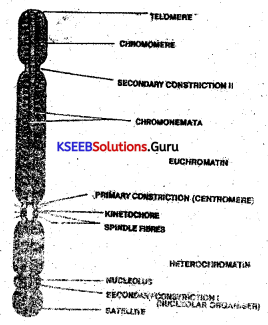
General structure of a Chromosome:
A typical structure of the chromosome under light microscope reveals the following details:
1. Centromere: It is also called primary constriction or Kinetochore that encloses Kinesomes that are joined together by chromatin fibrillae. It is a non-fixed constriction that holds the arms of the chromosome together and it is a part at which the spindle fibres attach to mobilize the chromosome during cell division.
Chromatid: It is called the arm of the chromosome that encloses a zig-zag arrangement of thread like structures called chromonema.
Upon the chromonema, there are bead like structures called chromomeres which are a collection of genes being concentrated upon the same locus. The chromonema and the chromomere are normal in their activity due to the presence of a matrix or ground substance that contains metallo-proteins, RNA, DNA, salts and traces of water.
One end of the chromatid shows a satellite body and such a chromosome is called a sat-chromosome. The other end is a non sticky telomere end.’ The satellite body is believed to help the movement of genes during gene recombination.
Note:
Euchromatin and Heterochromatin: Euchromatin refers to less coiled region of chromatin. It is less stained and contain genes which are active. Heterochromatin refers to the chromatin which is highly coiled. It takes more stain and contain genes which are less active or inactive.
Types of Chromosomes:
I. Classification of chromosomes based on the number of centromeres:
(a) Acentric chromosome: It is a chromosome without centromere.
(b) Monocentric chromosome: Here there is one centromere to hold the chromatids together.
(c) Bicentric chromosome: It is the presence of two centromeres in a chromosome.
(d) Polycentric chromosome: It is more than three centromeres in a chromosome.
![]()
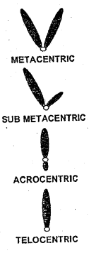
II. Classification of chromosomes based on the position of centromere:
(a) Metacentric type
Here the centromere is exactly at -the centre of two chromatids, It looks Y – shaped during anaphase,
(b) Sub metacentric type
Here the centromere is eccentric in position so that one of the chromatid is long and the other is shorter. It looks L – shaped ‘ during anaphase.
(c) Acrocentric type
Here the centromere is almost towards one end of chromatid to form a very long arm and another very short arm. It looks hook shaped during anaphase.
(d) Telocentric type
Here the centromere is towards one end of chromatid such that one chromatid is only present. It looks rod shaped during anaphase.
III. Classification of chromosomes based on their functions:
(a) Autosomes[AA]: They are also called somatic chromosomes that control the body characteristics.
(b) Allosomes [X or Y]: They are also called sex chromosomes that determine the gender of an individual.
Functions of chromosomes:
1. Chromosomes are very important in the higher animals for the phenomenon of sex determination.
2. Chromosomes play an active role in the metabolic process of a cell.
3. They carry the heredity information from parents to offsprings in the form of genes.
Ribosomes:
Ribosomes are asymmetrical particles which are dense, granular structure found attached to Endoplasmic reticulum or found floating freely in the cytoplasm. Also they are present in mitochondrial matrix and stroma of chloroplast. They are the sites of protein synthesis. They are present in both prokaryotes and in eukaryotes.
Chemically they contain 40% proteins and 60% rRNA and their number in a cell varies from 10,000 to millions. Based on the sedimentation co-efficient i.e., how fast a cell organelle sediments in an ultracentrifuge and expressed in swedberg unit there are two types of ribosomes, namely 70s seen in prokaryotes, plastids and mitochondria and 80s – seen in eukaryotes.
Each ribosome has two sub units, one is larger than the other. 70s ribosome has a large unit 50s and a small unit of 30s and 80s has the larger unit 60s and smaller unit 40s. Generally ribosomes are found in large groups called polyribosomes.
Functions:
1. Ribosomes are the sites of protein synthesis.
2. Free ribosomes produce enzymes for intercellular use. e.g: skin cells, erythroblasts.
3. Bound ribosomes synthesise enzymes for extracellular use. e.g: pancreatic cells,plasma cell etc.
![]()
Ergastic Substances:
The non-living inclusions of the cell are called Ergastic substances. These are either products of metabolism or by-products which are of varied nature. They occur in cytoplasm, vacuole or in cell wall. They may be classified into:
(a) Reserved food materials
(b) Secretory products
(c) Excretory products
(d) Mineral crystals
(a) Reserve food materials: The reserve food materials in the cell includes starch grains, protein grains, oil globules in plant cells and glycogen granules in the animal cell.
(b) Secretory products: These are chemical compounds secreted by the cytoplasm or cell organelles. They include enzymes, hormones, pigments like chlorophylls, carotene and xanthophylls in plants and haemoglobin in animals and Neetor (produced by nectaries in flowers).
(c) Excretory products: Most of the waste products are formed due to metabolic activities in the plant body and are commercially important materials. They accumulate in leaves, fruits, bark and sometimes in the roots of the plants. They include
(a) Alkaloids: present in roots, bark, leaves and fruits of plants.
e.g: Caffeine in coffee seeds, Theanine in tea leaves, nicotine, morphine, atrophin, azadiractin etc.
(b) Essential oils: These are present in leaves (lemon), skin of fruits (orange), flowers (rose jasmine) etc. .
(c) Resins: Present in resin ducts of the stem of conifers like pinus. Resins are used in biological preparations like microslides.
(d) Gums: They are liberated from many plants like Acacia, Neem plant etc., they are liberate through the cut ends.
(e) Latex: It is a milky, water soluble substance produced by laticiferous tissue in plants like euphorbia, calatropis, ficus etc.(Rubber).
(f) Tanins: They are polyphenols found in bark, leaves, stems and fruits of certain woody , plants Tanins are used for leather tanning.
(g) Mineral Crystals: The inorganic materials in cytoplasm many times accumulates in the – form of mineral crystals.
(h) Cystolith: It is a cluster of CaC03 present in palisade parenchyma of banyan leaf.
(i) Raphides: Crystals of calcium oxalate found in vacuoles. They are generally needle shape and occur in bundles as in Eichornia, Canna etc.
Vacuole:
These are non cytoplasmic areas, present inside the cytoplasm and bounded by a single layer of semipemfeable membrane called Tonoplast. Vacuoles mainly occur in Eukaryotes. In animal cells, the size of the vacuole is smaller and in plant cells, the size is larger and is central in position.
![]()
Functions:
Based on functions, vacuoles are of the following types:
(a) Sap vacuoles: Present in plant cells which store water and dissolved mineral ions. They function as osmometers.
(b) Contractile vacuoles: Present in protozoans like amoeba, paramaecium etc., help in osmoregulation and excretion.
(c) Food vacuoles: Present in protozoans and help in the digestion of food substances.
(d) Gas vacuoles: Present in protozoans,-contain gases like O2, CO2, N2 etc., and provide buoyancy.